Fig. 3.
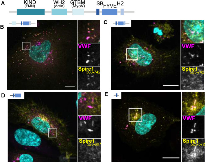
The SB domain of Spire1 is sufficient for mediating recruitment to WPB. A Depicts the domain structure of Spire 1 consisting of an actin-binding (WH2) and formin recruiting (KIND) N-terminal part. The C-terminal part comprises a Spire box domain (SB), a FYVE-type zinc finger and a H2 sequence similar to Slp/Slac proteins. B–E HUVEC were transfected with different GFP-tagged Spire1 truncation mutants (yellow) and co-stained for VWF (magenta). DAPI stainings are shown in cyan. Scale bars: 10 µm. The boxed areas are shown enlarged as a merge (upper picture) and as single channels (lower pictures, VWF and Spire mutants) next to the overview picture showing the whole cell. B Spire1 amino acid (aa) 388–742 (GTBM-SB-FYVE-H2, Spire1Tail). C Spire1 aa 525–742 (SB-FYVE-H2). D Spire1 aa 525–697 (SB-FYVE). E Spire1 aa 525–572 (SB)

