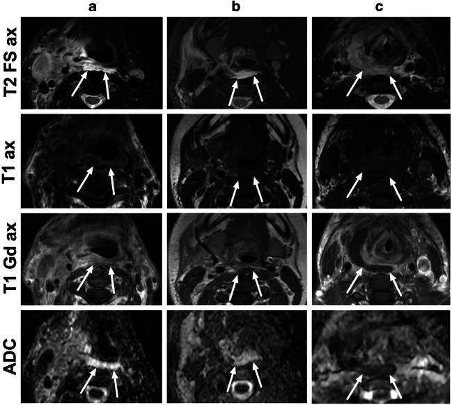
Fig. 2.
Different types of RPE in axial fat-suppressed T2-weighted Dixon images (first row), axial pre-contrast T1-weighted images (second row), axial post-contrast in-phase Dixon T1-weighted images (third row), and axial ADC maps (fourth row), from three patients (a–c). a A 63-year-old male with tonsillitis had high T2 signal, contrast enhancement on T1, and no purulence on ADC (enhancement-type RPE). b A 23-year-old female with parotitis had high T2-signal, non-enhancing fluid on T1, and no purulence on ADC (fluid-type RPE). c A 41-year-old female with a throat infection had an intermediate T2-signal, non-enhancing fluid on T1, and low ADC consistent with purulent fluid (true retropharyngeal abscess)

