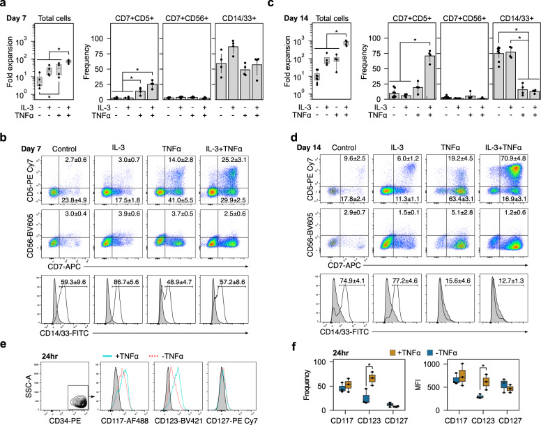
Fig. 3. IL-3 and TNFα enhance proT-cell expansion and purity.
CD34+ HSPCs were placed on DL4 + VCAM-1 and fold expansion and phenotype were measured on days 7 and 14. a Fold expansion and frequency of CD7+CD5+ proT, CD7+CD56+ NK, and CD14/33+ myeloid cells on day 7. Combining TNFα with IL-3 significantly increased total cell expansion over all other conditions. It also increased the frequency of CD7+CD5+ cells without increasing CD7+CD56+ frequencies. Box plots show median and interquartile range from n = 4 independent UCB donors and bar plots are mean ± standard error (*p < 0.05). b Representative flow cytometry plots on day 7. Frequencies are mean ± standard error. c By day 14, the frequency of CD14/33+ cells was significantly lower in groups containing TNFα than those without. Box plots show median and interquartile range from n = 4 independent UCB donors and bar plots are mean ± standard error (*p < 0.05). d Representative flow cytometry plots from day 14 show a relatively pure population of CD7+CD5+ cells when TNFα is combined with IL-3. Frequencies are mean ± standard error. e Representative flow cytometry plots of CD117, CD123, and CD127 expression on CD34+ HSPCs with or without TNFα stimulation for 24 h. f TNFα induced a significant increase in the frequency of CD123+ cells. The increased frequency of CD123+ cells was accompanied by an increase in the median fluorescent intensity (MFI) of CD123, indicating a higher receptor density on the cell’s surface after TNFα stimulation. *p < 0.05 for n = 3 independent UCB donors.

