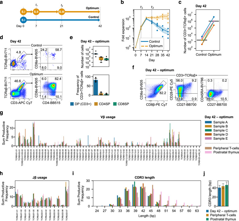
Fig. 5. Optimized cytokine concentrations provide stage-specific signals during T-cell development.
a The three-stage optimum cytokine concentrations were compared to a control that used the first stage concentrations throughout the entire differentiation. All other parameters (seeding density, passage ratio, schedule, etc.) were kept the same for each condition. b Fold expansion over 42 days using the optimized three-stage assay. Cytokine concentrations were changed at t1 and t2. Lines represent the mean over time for each condition and the shaded areas are the 95% confidence intervals. c Absolute count of CD3+TCRαβ+ cells on day 42 from 2000 HSPCs seeded per well on day 0. Lines connecting control and optimum are from the same UCB donor and the horizontal line is the median for each condition. d On day 42, CD3+TCRαβ+ cells were predominantly DP with some CD4SP and CD8SP T-cells present. e Absolute count and frequency of CD3+ DP, CD4SP, and CD8SP T-cells on day 42. The number and frequency of CD4/8 SP cells were comparable at this timepoint. The box plot shows median and interquartile range while the bar plot shows mean ± standard error. f On day 42, CD3+TCRαβ+ cells generated using the optimized cytokines were predominantly CD8αβ+ and expressed CD28. A small population of CD28+CD27+ cells were present and no cells expressed CD45RA. g Vβ gene diversity was comparable to peripheral T-cells and postnatal thymocytes. h Jβ gene diversity followed similar patterns of recombination to peripheral T-cells and postnatal thymocytes. i CDR3 lengths were similar in differentiated T-cells as peripheral T-cells and postnatal thymus. j Mean CDR3 length was similar between T-cells differentiated using optimized cytokines, peripheral T-cells, and postnatal thymocytes. From n = 5 UCB donors. One donor each of peripheral T-cells and postnatal thymocytes were included for comparison.

