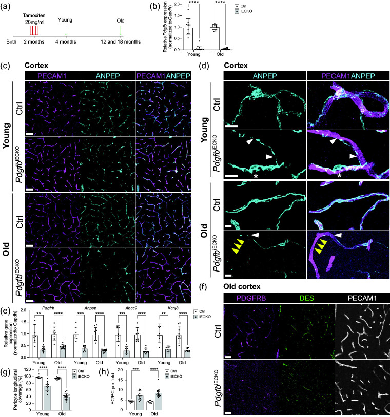
Figure 1.
Experimental scheme, gene deletion efficiencies and assessment of pericytes in adult-induced PdgfbiECKO and littermate controls. (a) Endothelial-specific Pdgfb deletion was accomplished by Tamoxifen administration for 5 days at 2 months of age. All analyzes were performed at 4 months of age for the “young” age and at 12- and 18-months for the “old” age. (b) qPCR analysis of Pdgfb mRNA expression on freshly isolated brain microvascular fragments. Pdgfb mRNA expression was normalized to endogenous Gapdh levels. In young mice, 8% of Pdgfb expression remained in PdgfbiECKO mice (n = 10). In old mice, 4% of Pdgfb expression remained in PdgfbiECKO mice (PdgfbiECKO n = 11, Ctrl n = 10). (c) Representative overview images of mural cells from the cortex of young and old mice. Co-immunolabeling of PECAM1 (magenta) and ANPEP (cyan). Scale bars 50 µm. (d) Representative high magnification images to visualize pericyte morphology in young and old PdgfbiECKO and controls. Co-immunolabeling of PECAM1 (magenta) and ANPEP (cyan). White arrowheads indicate fragmented pericyte processes. Asterisk indicates pericytes with altered cell bodies and distinct foot processes in PdgfbiECKO. Yellow arrowheads indicate shorter processes leaving part of the vasculature uncovered in PdgfbiECKO. Scale bars 10 µm. (e) qPCR analysis on the mural cell genes Pdgfrb and Anpep and the pericyte genes Abcc9 and Kcnj8 performed on freshly isolated brain microvascular fragments from young and old mice (for litter and n number see Supplementary Table 1). The genes of interest were normalized to endogenous Gapdh levels and are presented as relative gene expression to Ctrl samples. (f) Representative overview images from the cortex of old mice. Co-immunolabeling of PDGFRB (magenta), DES (green) and PECAM1 (white). Scale bars 25 µm. (g) The skeletal length of PECAM1 positive capillaries and ANPEP positive pericytes in PdgfbiECKO and controls was measured and plotted as the percentage of pericyte longitudinal length over blood vessel length. Three litters were analyzed for pericyte coverage in the cortex of young mice (PdgfbiECKO n = 11, Ctrl n = 8) and five litters were analyzed for coverage in the cortex of old mice (PdgfbiECKO n = 16, Ctrl n = 13). (h) Quantification of endothelial cell (ERG+) to pericyte (ANPEP+, DAPI+) ratio per field in young (PdgfbiECKO n = 11, Ctrl n = 8) and old mice (PdgfbiECKO n = 16, Ctrl n = 13). b, g and h-Old, normality tests revealed that the data was unevenly distributed so nonparametric Mann-Whitney U test was used to evaluate significance. e and h-Young, the significance of evenly distributed data was evaluated using unpaired 2-tailed t test with Welch’s correction. e, Gene expression comparison between young and old PdgfbiECKO mice was not significant for neither of the four genes. Data is presented as geometric mean with geometric SD. **p < 0.01, ***p = 0.001, ****p < 0.0001, ns = not significant.

