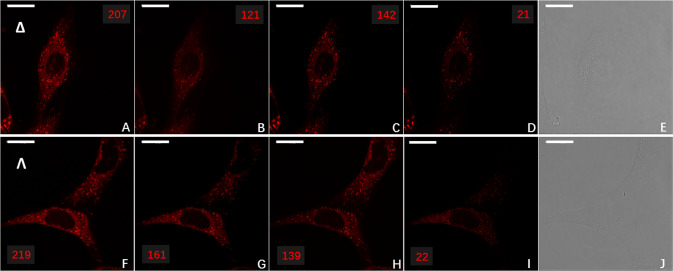
Fig. 5. Enantioselective localisation of enantiopure europium complex, Eu:L1 to the lysosome or mitochondria in live NIH 3T3 cells.
Enantioselective differential chiral contrast (EDCC) CPL-LSCM of Eu:L1 (30 µM, 14 h loading, ×63 1.4 NA oil objective, 96 × 96 µm FOV, 100 avg., 790 nm axial section) in live mouse skin fibroblast (NIH 3T3) cells, showing enantioselective localisation to the lysosome (Δ-Eu:L1) and mitochondria (Λ-Eu:L1). Top row Δ-Eu:L1. A Total Europium emission (λex = 355 nm, 20 mW, λem = 589–720 nm). B, C Left and Right CPL channel (λex = 355 nm, λem = 589–599 nm), respectively. D Left-handed EDCC image (left CPL—right CPL) highlighting enantioselective predominantly lysosomal localisation. E Transmission image. Bottom row Λ-Eu:L1. F, G, H, J as per (A, B, C, E). I Left-handed EDCC image (left CPL—right CPL) highlighting enantioselective predominantly mitochondrial localisation. Scale bars = 20 µm, numbers in red are avg. Eight-bit pixel intensity values for each image region.

