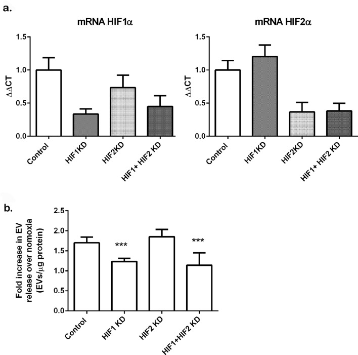
Figure 4.
Knockdown of HIF1α reduces EV release in hypoxic HEK293T cells. Cells were transfected with plasmids encoding for validated shRNAs specifics for HIF1α, HIF2α or both as detailed in the methods section. Following selection of transfectants in puromycin, cells were exposed to normoxic or hypoxic conditions for 16 h. (a) Expression of HIF1 and HIF2 by real time PCR. Total RNA was isolated from cells and mRNA abundance for HIF1α or HIF2α was determined by real time PCR, Data is mean ± SDEV of a representative experiment, of N = 4 experiments. (b) EVs were isolated by differential centrifugation protocol after an ultracentrifugation step at 100,000×g for 75 min to pellet EVs. EVs were analysed by NTA. Data shows mean ± SEM of N = 3–4 experiments per group. EVs were normalized to total protein content in the cellular lysates and referred to the normoxic group to combine the data from different experiments. Statistical analysis One Way ANOVA: ***indicates p ≤ 0.05.

