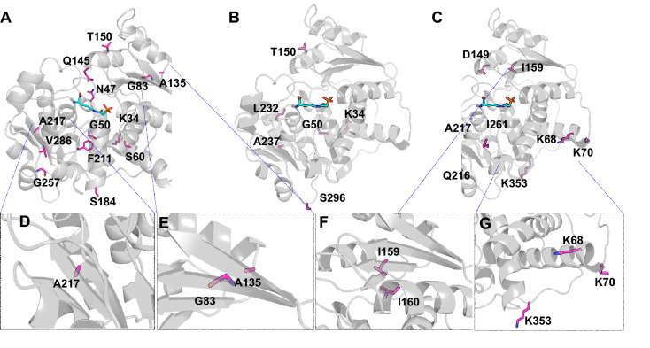
Figure 7.
Structural mapping of important individual residues onto hOTC monomeric structure. A structure of the hOTC monomer (white cartoon) bound to a substrate analog (N-phosphonoacetyl-L-ornithine, cyan sticks) (PDB: 1OTH) is used to highlight amino acid positions mutated in the VAE-sampled sequences (magenta sticks). (A) Amino acid positions of mutations that have a beneficial/positive effect on Tm and specific activity. (B) Amino acid positions of mutations that have a beneficial/positive effect on either Tm or specific activity, but not both. (C) Amino acid positions of mutations that negatively affect Tm and specific activity. (D–G) Close-up views of A217, A135 and G83, the small beta-sheet consisting of I159 and I160 and the three important lysines at K68, K70 and K353.

