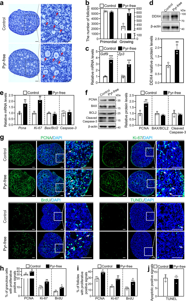
Fig. 2. Effect of short-term pyruvate deprivation on mouse primordial follicle activation in vitro.
Ovaries at 2 dpp were cultured in standard (control) or pyruvate-free (pyr-free) medium for 1 day (e–j) or 2 days (a–d). a, b Morphological comparison of the ovaries (a) and the number of primordial and growing follicles (arrows, b) in the control and pyruvate-free group. Nuclei were stained by hematoxylin. Scale bars: 50 μm. c The mRNA levels of Gdf9 and Zp3 in the control and pyruvate-free group. d DDX4 protein levels in the control and pyruvate-free group. e The mRNA levels of Pcna, Ki-67, Bax/Bcl-2, and Caspase-3 in the control and pyruvate-free group. f The protein levels of PCNA, BAX, BCL-2, and Cleaved Caspase-3 in the control and pyruvate-free group. g Immunofluorescence stain of PCNA, Ki-67, BrdU, and TUNEL (green) in the control and pyruvate-free group. DAPI, blue. Scale bars: 50 μm. h–j The percentage of granulosa cells (h) and primordial follicles (i) with PCNA-, Ki-67-, or BrdU-positive signals, and the number of cells with TUNEL-positive signals (j) in the control and pyruvate-free group. All the experiments were repeated three times, and the representative images are shown. In western blot results, β-actin was used as an internal control. Bars indicate the mean ± SD. *P < 0.05, **P < 0.01, and ***P < 0.001 vs. control.

