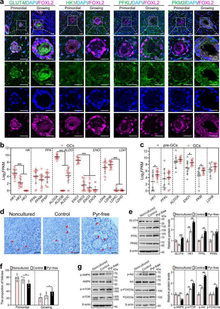
Fig. 6. Effect of short-term pyruvate deprivation on human primordial follicle activation in vitro.
a Immunofluorescence stain of GLUT4, HK1, PFKL, and PKM2 (green) in human primordial and growing follicles. FOXL2, purple; DAPI, blue. Scale bars: 20 μm. b, c Log2FPKM values were extracted from previously published data (GSE107746). The expression levels of isoforms in each family of HK, PFK, ALDO, ENO, and LDH in human granulosa cells of primary follicles (n = 15 follicles, b), and the expression levels of HK1, PFKL, ALDOA, ENO1, PKM, and LDHB in human granulosa cells of primordial (n = 8 follicles) and primary follicles (n = 15 follicles, c). GCs granulosa cells, pre-GCs pre-granulosa cells. d–g Human ovarian fragments were directly fixed in 4% PFA (noncultured), or cultured in standard medium (control), or cultured in the pyruvate-free medium for 2 days and then in standard medium for indicated days (pyruvate-free group). The fragments were collected after 3 days (e, g) and 6 days (d, f) of treatment, respectively. d, f Morphological comparison of human ovarian tissue fragments (d) and the proportion of primordial (arrowheads, f) and growing follicles (arrows, f) in the control and pyruvate-free group. (n = 5 independent experiments). Nuclei were stained by hematoxylin. Scale bars: 50 μm. e The protein levels of GLUT4, HK1, PFKL, and PKM2 in the noncultured, control, and pyruvate-free groups. g The protein levels of p-AMPK, p-mTOR, p-Akt, and p-FOXO3a in the noncultured, control, and pyruvate-free groups. The representative images are shown. In western blot results, the levels of total AMPK, mTOR, Akt, and FOXO3a were used as the corresponding internal control for p-AMPK, p-mTOR, p-Akt, and p-FOXO3a, respectively, and β-actin was used as the internal control for GLUT4, HK1, PFKL, and PKM2. Bars indicate the mean ± SD. *P < 0.05, **P < 0.01, and ***P < 0.001.

