Fig. 1.
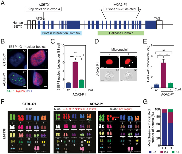
SETX deficiency promotes chromosome fragility. (A) Diagram of human SETX showing ΔSETX and the AOA2-P1 mutant fibroblasts. Boxes represent exons and the positions of start (ATG) and stop (TAG) codons. (B) Immunostaining with 53BP1 (green) and cyclin A (red) antibodies in control (CTRL-C1) and AOA2-P1 fibroblasts. DNA was stained with DAPI (blue). Representative images of G1- and S-phase cells are shown. (Scale bars, 5 μm.) (C) Quantification of 53BP1 NBs in G1 cells, as in B, with or without cordycepin. Data represent the mean ± SEM of three independent experiments with >300 cells per condition. (D) Imagestream imaging flow cytometry of AOA2-P1 fibroblasts. Nuclei were stained with PI. Representative PI and bright field (BF) images of mononucleated cells with and without micronuclei are shown. White arrows denote micronuclei. (Scale bars, 7 μm.) (E) Quantification of cells with micronuclei, as in D. Cells were treated with or without cordycepin. Data represent the mean ± SEM of three independent experiments, >4,000 cells per condition. (F) M-FISH analyses of metaphase spreads from CTRL-C1 and two AOA2-P1 fibroblasts showing deletions/amplifications and chromosome fragility. Representative karyotypes are shown. White arrows, chromosome aberrations. (G) Quantification of metaphases with indicated number of aberrations, as in F. Thirty metaphases were analyzed per condition. **P < 0.01 and ****P < 0.0001 by Mann–Whitney U test. P ≥ 0.05 is considered not significant (ns).

