Fig. 1.
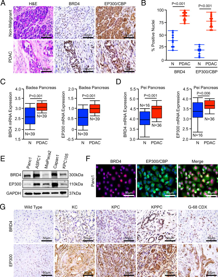
BRD4 and EP300/CBP are ubiquitously expressed in PDAC. (A and B) Excisional biopsies from 14 PDAC patients, nine with matched, adjacent nonmalignant tissue, were sectioned and stained for either BRD4 or EP300/CBP. The number of positive nuclei per 40× field was quantified by three blinded investigators and divided by the total number of nuclei in each field. These values were averaged and displayed as an individual value plot. (C and D) Pancreatic tumor tissues (PDAC) and adjacent nonmalignant (N). The Badea et al. and Pei et al. cohorts of PDAC patients were evaluated for mRNA expression of BRD4 or EP300 using the Oncomine platform. All mRNA expression values are plotted in log scale. (E) Human PDAC cell lines Panc1, ASPC1, MiaPaCa2, Capan1, and the murine PDAC cell line KPC-105 were evaluated for expression of BRD4 and EP300/CBP by Western blot. (F) Panc1 cells were stained for BRD4 and EP300/CBP by immunocytochemistry. (G) Pancreas tissue from either nongenic wild type, the KC model of PanIN disease, the KPC model of advanced PDAC, the KPPC model of extremely aggressive PDAC, or subcutaneous tumor tissue from the G-68 CDX model were collected and stained for either BRD4 or EP300/CBP.

