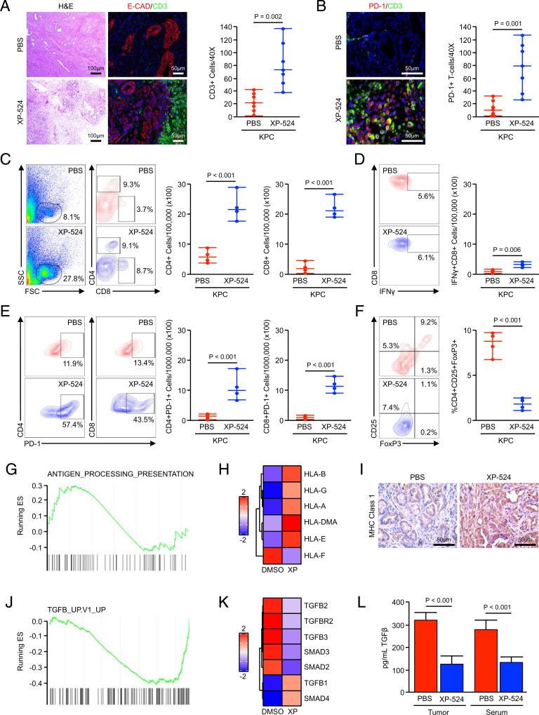
Fig. 6.
XP-524 enhances T cell recruitment but fails to promote a functional antitumor immune response. (A and B) KPC mice were generated as a model of advanced PDAC. Starting at 15 wk of age, mice were administered daily IP injections of either PBS vehicle or 5 mg/kg XP-524. Pancreas tissues were collected when the animals were moribund and stained with either H&E or via IHC for E-cadherin and the T cell surrogate CD3 or for CD3 and T cell exhaustion marker PD-1. Tissue sections were quantified as described and displayed as an individual value plot. (C) KPC were enrolled as described and treated with either a PBS vehicle or 5 mg/kg XP-524. After 2 mo on treatment, tumor tissues were collected and analyzed by flow cytometry for tumor-infiltrating CD4+ and CD8+ T cells, respectively (n = 4 per group). FSC, forward scatter; SSC, side scatter. (D) Tumor-infiltrating cells were gated based on the CD8 staining shown previously, and the total number of cells positive for IFN-γ were displayed as an individual value plot. (E) The total number of cells positive for either CD4 or CD8 and T cell exhaustion marker PD-1 from the tumors of PBS- and XP-524–treated mice. (F) Tumor-infiltrating T cells were gated based on CD4 staining and analyzed for CD25 and FoxP3, and the total number of CD4+CD25+FoxP3 Tregs were displayed as individual value plot. (G and H) Panc1 cells were treated with either a DMSO vehicle or 1 μM XP-524, analyzed by RNA sequencing, and subjected to GSEA revealing a significant enrichment for the antigen-processing and presentation pathway. (I) Tissues from KPC mice treated with either PBS or XP-524 were stained via IHC for MHC Class 1 and representative images displayed. (J and K) RNA sequencing and GSEA for Panc1 treated as described, revealing a significant down-regulation of the TGFβ signaling pathway. (L) Homogenized tumor tissue or serum from KPC mice treated with either PBS or XP-524 were analyzed for levels of TGFβ1 by ELISA.

