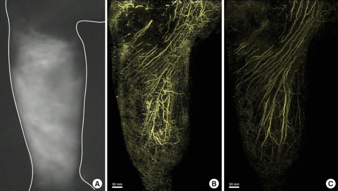
Fig. 2.

Lymphedema of the right leg after surgery for lymphoma. An 81-year-old man presented with lymphedema of the right leg after surgery, including pelvic lymph node dissection for lymphoma. An image of the medial side of the right lower leg examined using indocyanine green fluorescence lymphography shows dermal backflow in the entire lower leg (A). A photoacoustic lymphangiography image shows the lymphatic capillaries of the network structure in the superficial layer and a linear appearance of the lymphatic collectors of the deep layer (B). A photoacoustic lymphangiography image of the deep layer shows the lymphatic collectors (C).
