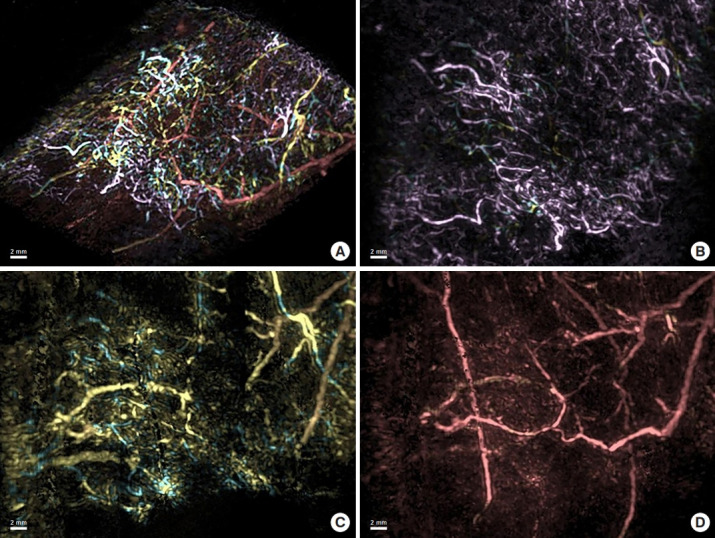
Fig. 5.

Three-dimensional image of dermal backflow. A superimposed image on a tilted plane of the three-dimensional structure of dermal backflow is shown with the superficial vessels in pink, the intermediate layer vessels in yellow, and the deep vessels in red (A). Each layer in the sagittal plane is shown individually (B, C, D). Capillary lymphatics are shown in the dermis (pink; B), precollectors are shown in the deep layer of the dermis (yellow; C), and lymphatic collectors are shown in the subcutaneous tissue (red; D).
