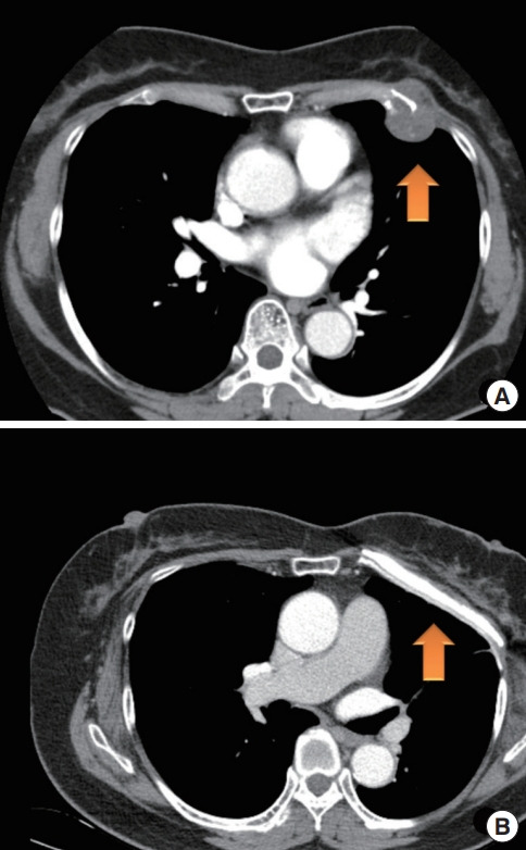
Fig. 3.

Preoperative and postoperative computed tomography (CT) images showing the chest wall. (A) Preoperative CT shows chondrosarcoma on the patient’s left fourth rib cartilage (arrow). (B) At 11 months after surgery, postoperative CT shows three layers of the reconstructed chest wall (arrow).
