Figure 5.
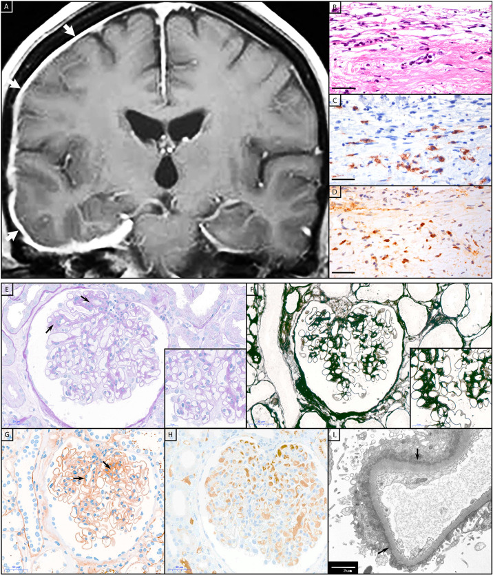
Histopathology of IgG4-RLD and IgG4-AID patients. Patient with IgG4-RLD: (A) MRI shows a right-sided thickening and increased contrast agent uptake of the pachymeninges (white arrows). A biopsy from the meninges reveals fragments of dura with (B) prominent fibrosis and infiltration with (C) numerous CD138+ and (D) IgG4+ plasma cells, compatible with IgG4-related pachymeningitis. Patient with IgG4-AID: A kidney biopsy from a patient with CNTN1/Caspr1-complex autoantibodies and acute kidney failure, nephrotic syndrome, hypoalbuminemia and microhematuria. (E) Glomerulus with mild segmental fibrous mesangial expansion (arrows), somewhat thickened capillary basal membranes, without hypercellularity (PAS stain). (F) Silver stain with smooth capillary loop basal membranes. (G) Immunohistochemistry for IgG with global finely granular peripheral and segmental mesangial positive deposits (arrows). (H) Negative immunohistochemistry for IgG4. (I) Electron microscopy with numerous small sub-epithelial electron-dense deposits with flattening of podocytic foot processes (arrows). Scale bar (B–D), (E–H) = 50 µm; scale bar inset in (E, F) = 20 µm; scale bar (I) = 2 µm.

