
FIGURE 1.
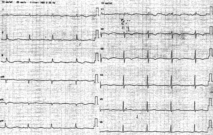
ECG on admission shows a HR of 48 beats/min, no axial deviation, complete right bundle branch block, I‐degree AV block I, II, aVR, aVL, aVF, V1‐6 negative T waves, flat and low T waves, biphasic changes, and a QTc of 480 ms (Bazett correction)