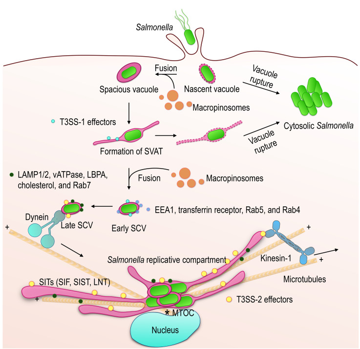
Figure 1. FIGURE 1: Maturation of the Salmonella-containing vacuole.
Salmonella is able to infect non-phagocytic cells through the activity of effectors injected into the cell by T3SS-1. The formation of membrane ruffles allows entry into the cell and induces the formation of macropinosomes. The nascent vacuole is weakened by the presence of T3SS-1 and prone to rupture (5–20% of bacteria). Salmonella replicates rapidly in the cytosol of epithelial cells. Fusion of the nascent vacuole with macropinosomes increases its volume (spacious vacuole) and stabilizes it. Spacious vacuoles undergo further membrane remodeling, including the formation of SVAT with the help of the T3SS-1 effectors (south sea blue dots) and the host proteins SNX1/3. SVAT formation promotes vacuole rupture if membrane loss is not compensated by fusion activity with macropinosomes. The Early SCV is characterized by the presence of early endosomal markers (light blue dots). Its maturation is marked by the loss of early endosomal markers, the acquisition of late endosomal markers (dark green dots) and a shift of the SCV from the cell periphery to the MTOC region. Changes in the content and physico-chemical properties of the SCV, in particular the drop in its pH, induce the expression of T3SS-2 and its effectors (yellow dots). These allow the establishment of the replicative compartment whose membrane is very similar to that of the lysosomes. During the replication phase, the SCVs remain in the juxtanuclear region and are associated with membrane tubules (SITs), of the same composition, that stretch into the cell, supported by the microtubules and under the action of associated molecular motors.

