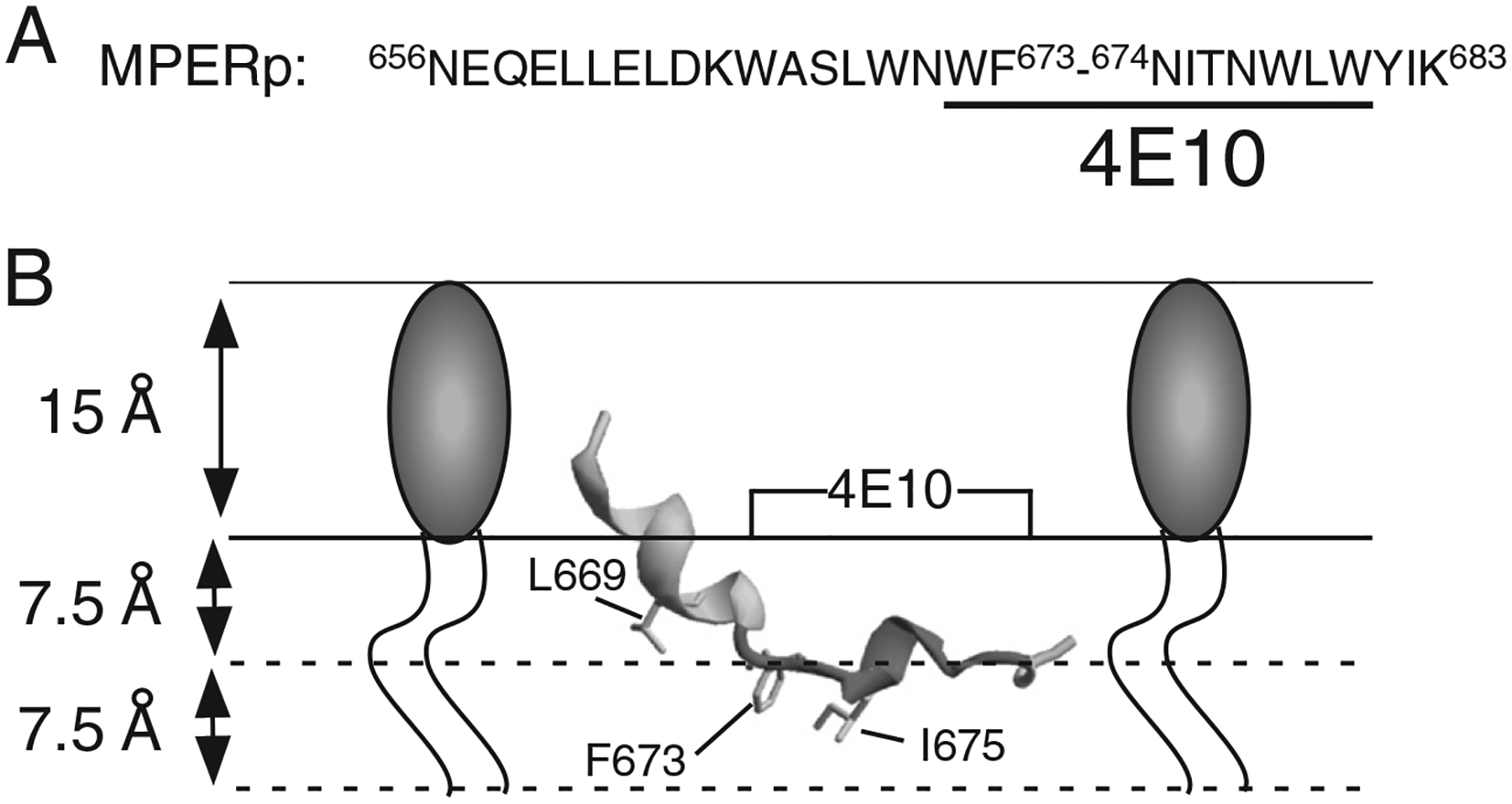
Fig. 1.

HIV MPER designation and model for its association with membranes Panel A: sequence of MPER peptide used in this study. 4E10 epitope residues underlined. Numbering is based on the prototypic HXBc2 viral isolate. Panel B: model for the cognate ELDKWASLWNWFNITNWLWYIK peptide in association with a membrane monolayer. The structure adopted in detergent micelles was obtained from the Protein Data Bank (PDB ID: 2PV6) and rendered using Swiss-PDB-viewer. The insertion depths for the depicted residues L669, F673 and I675 are based on electro paramagnetic spectroscopy determinations [12].
