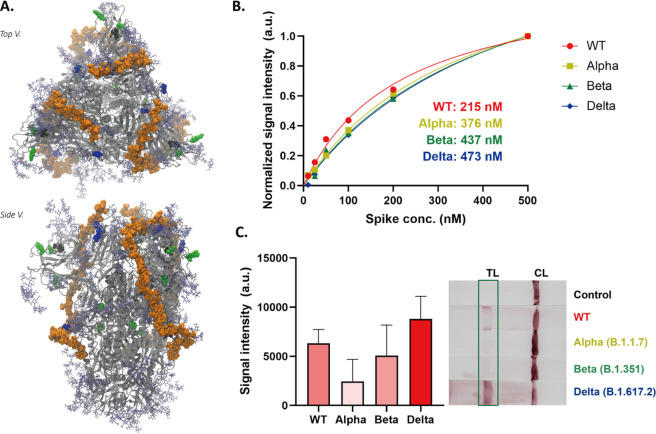
Figure 7.
(A) Trivalent hep40mer aligned to the Delta spike variant. Light green van der Waals spheres represent single point mutation sites, black van der Waals spheres represent locations of NTD deletion sites, and blue van der Waals spheres represent mutations along the hep40mer binding site L452K, T478R, and P681R. (B) BLI results of the HEP to spike proteins (wild type, Alpha (B.1.1.7), Beta (B.1.351), and Delta (B.1.617.2)). (C) Response of the Alpha, Beta, and Delta variants in the GlycoGrip LF biosensor. Statistical analysis was performed using one-way ANOVA with Tukey’s post hoc test.

