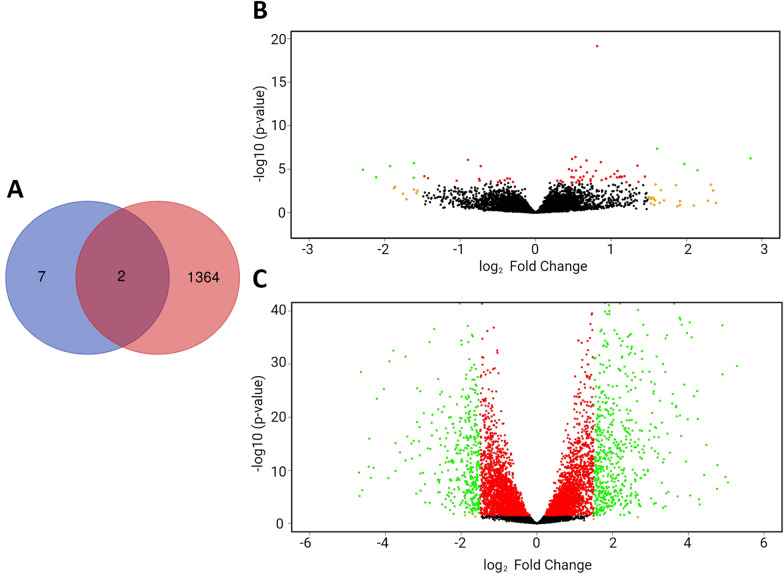
Figure 3.
Venn diagram and volcano plots of differentially expressed (DE) genes in protoscoleces from cattle and sheep liver and lung CE cysts. A Venn diagram showing the number of DE genes for each group compared. The number of DE genes in PSC from sheep liver and lung CE cyst samples (9 genes) are in purple. The number of DE genes in PSC from cattle and sheep liver CE cyst samples (1366 genes) are in red. Two genes are shared between both comparisons. B Volcano plot of DE genes. Each dot is a gene. Negative values belong to PSC from sheep lung CE cyst samples, positive values belong to PSC from sheep liver CE cyst samples. Green dots: FDR < 0.05 and log2FoldChange > 1.5, Orange dots: FDR > 0.05 and log2FoldChange > 1.5, Red dots FDR > 0.05 and log2FoldChange < 1.5. Black dots do not fit any of these criteria. C Volcano plot of DE genes. Each dot is a gene. Negative values belong to PSC from sheep liver CE cyst samples, positive values belong to PSC from cattle liver CE cyst samples. Green dots: FDR < 0.05 and log2FoldChange > 1.5, Orange dots: FDR > 0.05 and log2FoldChange > 1.5, Red dots FDR > 0.05 and log2FoldChange < 1.5. Black dots do not fit any of these criteria.

