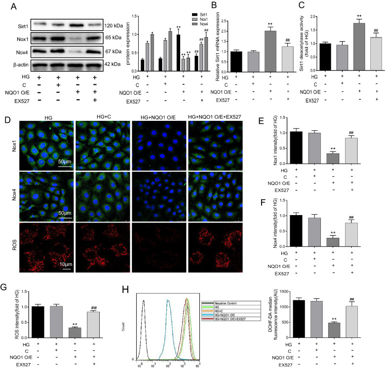
Fig. 7.
Effect of EX527 on oxidative stress in NQO1 pcDNA-treated HK-2 cells under HG conditions. A The protein levels of Sirt1, Nox1 and Nox4 were analyzed by western blotting (n = 3). B The mRNA level of Sirt1 was measured by RT-qPCR (n = 3). (C) Sirt1 activity in HK-2 cells (n = 6). D–F The expression levels of Nox1 and Nox4 were measured by immunofluorescence (scale bar, 50 μm, n = 6). D Mitochondrial ROS was assessed by the fluorescence probe MitoSOX red (Scale bar, 10 μm, n = 6). G The quantitative analysis of mitochondrial ROS. (H) Intracellular ROS was detected by flow cytometry and quantitative analysis was performed (n = 6). HG: 30 mM d‐glucose, HG + C: HG + control pcDNA3.1(+), HG + NQO1 O/E: HG + NQO1 pcDNA3.1(+), HG + NQO1 O/E + EX527: HG + NQO1 pcDNA3.1(+) + EX527 (1 μM). Values are expressed as the mean ± SD. **P < 0.01 versus HG + C group; ##P < 0.01 versus HG + NQO1 O/E group

