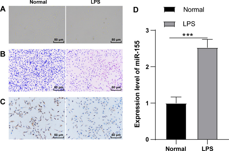
Fig. 1.
miR-155 was highly expressed in LPS-induced mouse KOA chondrocytes. The mouse knee chondrocyte inflammatory model was established by LPS induction. A The cell morphology was observed using an inverted microscope; B The cell morphology was observed using toluidine blue staining; C The expression of the knee chondrocyte marker Collagen II was detected by immunocytochemistry; D The expression of miR-155 was detected using RT-qPCR. The cell experiment was repeated 3 times. The data were expressed as mean ± standard deviation. Independent t test was used for data comparisons between 2 groups. *** P < 0.001

