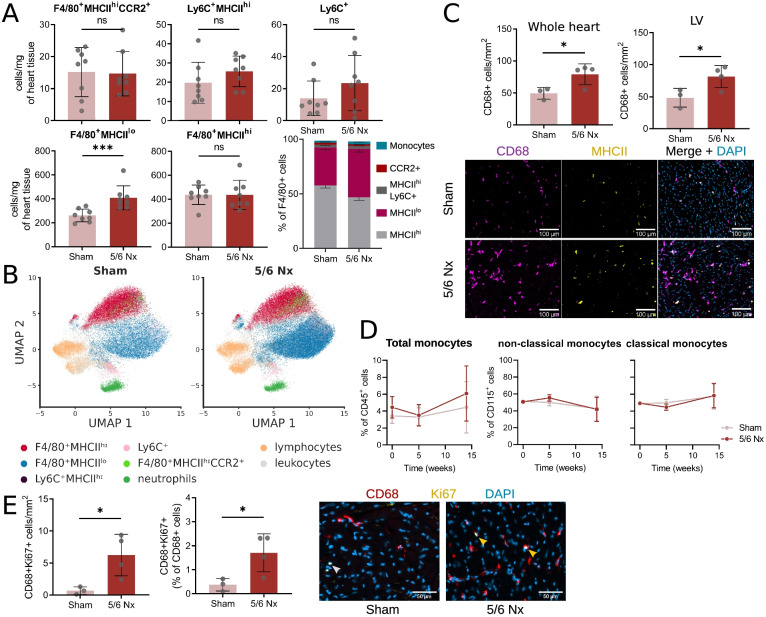
Fig. 3.
Cardiac macrophage numbers increase during chronic kidney disease. A F4/80+MHCIIhi and F4/80+MHCIIlo resident macrophage, Ly6C+MHCIIhi macrophage, F4/80+CCR2+MHCIIhi pro-inflammatory macrophage or Ly6C+ monocyte numbers per mg of heart tissue and proportion (stacked barplot) in sham and 5/6 nephrectomised (Nx) mice. (*** P < 0.001). B Representative parametric UMAP embedding for the visualisation of different leukocyte populations in sham (left) and 5/6 Nx (right) mice. C Number of CD68-positive macrophages per mm2 of cardiac tissue measured in whole heart slice and in the left ventricle (LV) and representative images showing CD68 MHCII co-staining (scale bar = 100 μm) in sham and 5/6 nephrectomised (Nx) mice (* P < 0.05). D Total blood monocyte (percentage of all leukocytes) and monocyte subset (non-classical and classical, as percentage of CD115+ monocytes) frequency over 12 weeks in sham and 5/6 Nx mice. Presented as percentage of all leukocytes (total monocytes) or Ly6C+ (classical monocytes) or Ly6C− (non-classical monocytes). Mean ± SD at each time point. n = 6. E) Proportion of Ki67-positive macrophages in CD68-positive macrophages measured in whole heart slice and representative images showing CD68 Ki67 co-staining (scale bar = 50 μm) in sham and 5/6 Nx mice

