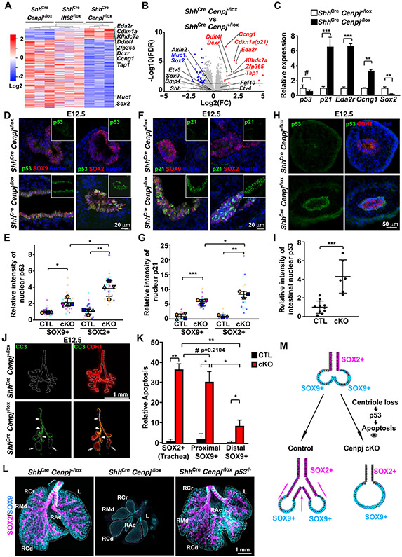
Figure 3. Centriole loss in endoderm activates p53-dependent apoptosis.
(A) Heatmap of E11.5 control (ShhCre Cenpj+/lox), Ift88 loss of function (ShhCre Ift88−/lox) and Cenpj loss of function (ShhCre Cenpj−/lox) lung RNAseq. Significant differentially expressed genes from Cenpj loss of function (ShhCre Cenpj−/lox) lung against control (ShhCre Cenpj+/lox), Ift88 loss of function (ShhCre Ift88−/lox) lungs were listed in heatmap. Cdkn1a (p21) and several other p53-induced genes are specifically upregulated upon loss of CENPJ.
(B) Volcano plots comparing log2 fold change of normalized RNAseq reads for E11.5 Cenpj loss of function (ShhCre Cenpj−/lox) as compared to control (ShhCre Cenpj+/lox) lungs. Dashed line denotes the p=0.05 cutoff. Selected genes upregulated ≥2 folds were indicated in red, and selected genes down-regulated ≥2 folds were indicated in blue (FDR <0.05).
(C) RT-qPCR measurement of Cdkn1a (p21), Ccng1, Eda2r, and Sox2 expression in E13.5 control (ShhCre Cenpj+/lox) and Cenpj loss of function (ShhCre Cenpj−/lox) lungs. Sox2 was downregulated and Cdkn1a (p21), Ccng1 and Eda2r upregulated upon loss of CENPJ. n=4, 4, 4, 4, 3, 3 respectively for the control groups; n=3, 4, 4, 4, 3, 3 respectively for Cenpj loss of function groups.
(D-E) Staining (D) and quantification (E) of p53 in E12.5 control (CTL, ShhCre Cenpj+/lox) and Cenpj loss of function (cKO, ShhCre Cenpj−/lox) lung cryo-sections. Scale bar, 20 μm. 4 independent replicates were analyzed for statistics.
(F-G) Staining (F) and quantification (G) of p21 in E12.5 control (ShhCre Cenpj+/lox) and Cenpj loss of function (ShhCre Cenpj−/lox) lung cryo-sections. Scale bar, 20 μm. 4 and 3 independent replicates for SOX9+ and SOX2+ respectively were analyzed for statistics.
(H-I) Staining (H) and quantification (I) of p53 in E12.5 control (ShhCre Cenpj+/lox) and Cenpj loss of function (ShhCre Cenpj−/lox) small intestine cryo-sections. Scale bar, 50 μm. n=10 and 6 for CTL and cKO respectively.
(J-K) Staining (J) and quantification (K) of cleaved Caspase3 (CC3, green) in E12.5 control (ShhCre Cenpj+/lox) and Cenpj loss of function (ShhCre Cenpj−/lox) lungs. Epithelia (CDH1, red) were outlined by dashed line. Arrows indicate the distal region of Cenpj loss of function lung, and arrowheads indicate the proximal part. Scale bar, 1 mm. n=3.
(L) Wholemount immunostaining of E15.5 control (ShhCre Cenpj+/lox), Cenpj loss of function (ShhCre Cenpj−/lox), Cenpj and p53 combined loss of function (ShhCre Cenpj−/lox p53−/−) lungs for SOX2 (magenta) and SOX9 (cyan). Scale bar, 1 mm.
(M) Schematic showing proposed mechanism by which centrioles participate in lung branching. In the absence of centrioles, SOX2-expressing, but not SOX9-expressing, cells activate p53 to trigger apoptosis.

