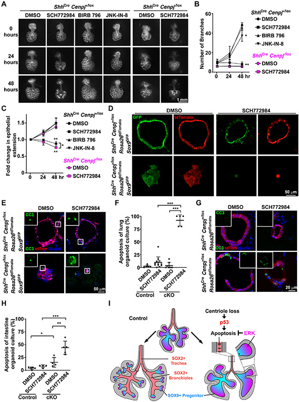
Figure 5. ERK activity protects acentriolar cells from p53-dependent apoptosis.
(A) Control (ShhCre Cenpj+/lox) and Cenpj loss of function (ShhCre Cenpj−/lox) lungs were isolated at E11.5 and cultured on a semi-permeable membrane. Control lungs were treated with DMSO, SCH772984, BIRB 796 or JNK-IN-8 for 48 hours. Cenpj loss of function lungs were treated with DMSO or SCH772984 for 48 hours. Images were taken every 24 hours. Scale bar, 1 mm.
(B-C) We measured the number of branches (B) and epithelial extension (C) of the lungs from (A). The fold change of epithelial extension was normalized to that immediately before the addition of the indicated compounds (i.e., at 0 hours). DMSO treatment was compared to BIRB 796, JNK-IN-8 or SCH772984 treatment within the same genotype using unpaired t-test. n=3.
(D) Visualization of GFP and tdTomato fluorescence in control (ShhCre Cenpj+/lox R26tdTomato Sox9GFP) and Cenpj loss of function (ShhCre Cenpj−/lox R26tdTomato Sox9GFP) lung organoid culture cryo-sections. Scale bar, 50 μm. Note: parameters of GFP fluorescence were adjusted in each group for better visualization.
(E-F) Staining (E) and quantification (F) of cleaved Casp3 (CC3) in control (ShhCre Cenpj+/lox R26tdTomato Sox9GFP) and Cenpj loss of function (ShhCre Cenpj−/lox R26tdTomato Sox9GFP) lung organoid culture cryo-sections. Inlets show the magnified view of cleaved Casp3 staining. Scale bar, 50 μm. DMSO treatment was compared to SCH772984 treatment within the same genotype, and different genotype with the same treatment was compared using unpaired t-test. n=6, 12, 8, 5 respectively.
(G-H) Staining (G) and quantification (H) of Cleaved Casp3 (CC3) stained in control (ShhCre Cenpj+/lox R26tdTomato) and Cenpj loss of function (ShhCre Cenpj−/lox R26tdTomato) intestinal organoid culture cryo-sections. Inlets show cleaved Casp3 staining. Scale bar, 20 μm. DMSO treatment was compared to SCH772984 treatment within the same genotype, and different genotype with the same treatment was compared using unpaired t-test. n=5, 4, 4, 5 respectively.
(I) Schematic illustrating the differential responses of lung epithelial cells to loss of centrioles. Loss of centrioles induced p53 activation in the endodermal epithelium. Acentriolar lung cells with low ERK activity (SOX2-expressing cells) apoptose, whereas acentriolar lung SOX9-expressing progenitors with higher ERK activity survive and grow.
See also Figure S5.

