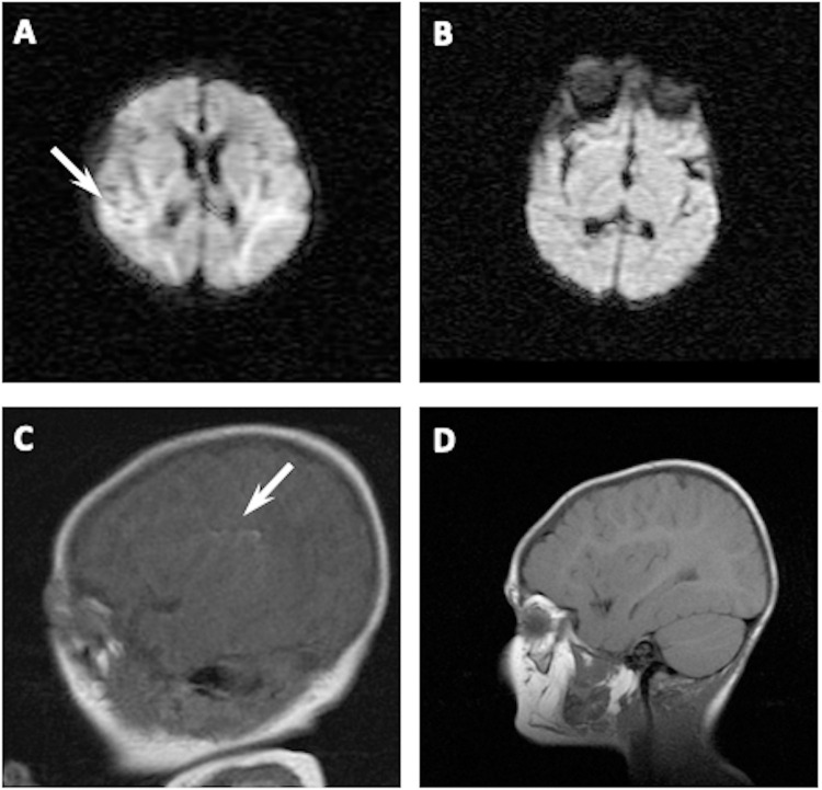
Fig 2. Representative MRI scans showing altered studies in two hypoxic-ischemic newborns.
In A, the MRI scan performed at postnatal day 5 in a female (39 weeks of gestational age) showed increased signal in both temporal lobes using Diffusion-Weighed Images; scan performed in the same patient aged 30 months showed no abnormalities (B). In C, the MRI scan performed at postnatal day 5 in a male (41 weeks of gestational age) showed linear hyperintensity in left insular subcortical area using T1-Weighed Images; scan performed in the same patient aged 26 months showed no abnormalities (D).

