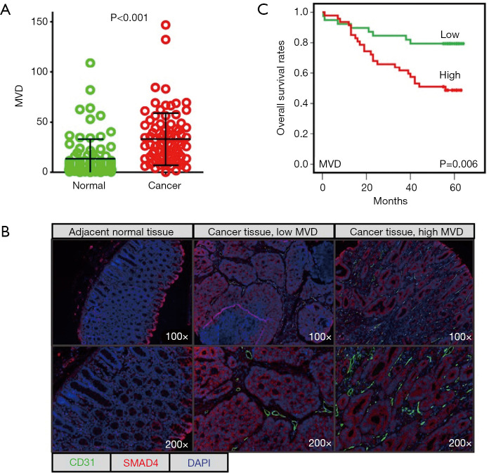
Figure 1.
mfIHC stains CD31 to show the microvessels in the tissues of colon cancer. (A) The MVD between cancer tissues and adjacent normal tissues were compared; (B) representative images show CD31-stained MVD for adjacent normal tissues, low MVD, and high MVD in cancer tissues. SMAD4 was stained to label the tumour cells; (C) Kaplan-Meier analysis of OS rates. mfIHC, multiplex fluorescent immunohistochemistry; MVD, microvascular density; OS, overall survival.

