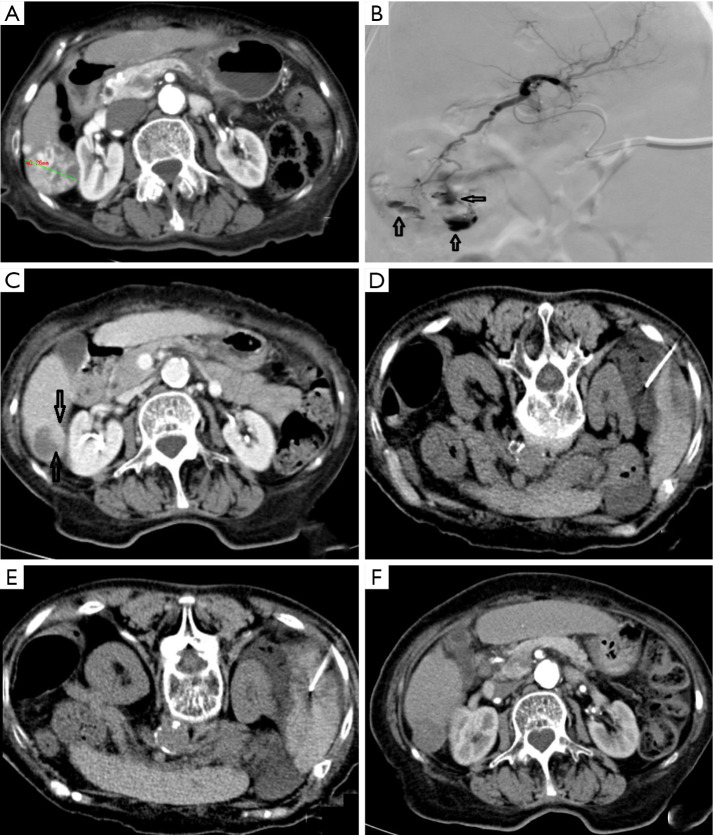
Figure 2.
A 75-year-old female received DEB-TACE for HCC. VL was found but not treated. Local residual tumors were treated with radiofrequency ablation. (A) Pre-operative CT scan revealed a 4.0-cm HCC lesion with rich blood supply in hepatic segment 6 (S6). (B) VLs (indicated by hollow arrows) were found during D-RACE. Stable VLs were considered, and no further treatment was applied. The embolization endpoint was SACE level III. (C) Local residual lesions (indicated by hollow arrows) around VL, along with necrotic tissues around the tumors, were found 2 months after operation. (D) After CT-guided puncture of the hepatorenal recess with a 21 G puncture needle, 500 mL of normal saline was injected to induce artificial ascites, which increased the distance between liver and kidney. (E) CT-guided radiofrequency ablation of residual lesions was performed using a 17 G radiofrequency needle, assisted by artificial ascites technique. (F) Two months after radiofrequency ablation, no residual tumors were found, the lesion area was not enhanced, and the lesion was reduced to 2 cm in diameter. DEB, drug-eluting beads; TACE, transarterial chemoembolization; HCC, hepatocellular carcinoma; SACE, subjective angiographic chemoembolization endpoint.

