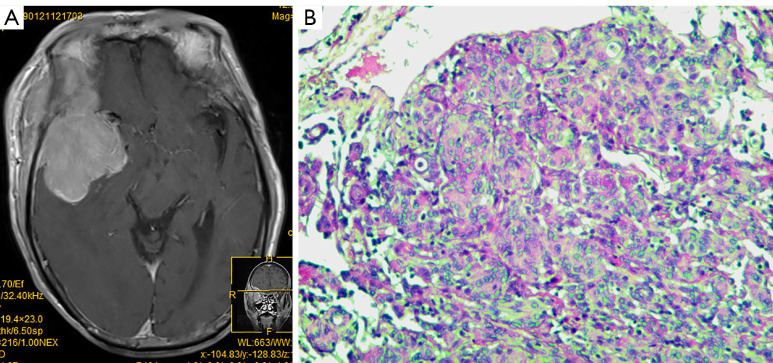
Figure 3.
MRI and pathological features of atypical meningiomas in clinical case 3. Male patient, 70 years old. (A) MRI shows a huge tumor with rich blood supply in the right temporal lobe, which has invaded the lateral wall of the right orbit causing sphenoid bone damage, and invasion of the infratemporal fossa, pterygopalatine fossa, and medial pterygoid and lateral pterygoid muscles outside the cranium. (B) HE staining (×100) shows tumor cells are arranged in small sheets, of papillary or whirlpool shape, with obvious nucleoli and abundant cytoplasm; Immunohistochemistry results show EMA (+/−), Vimentin (+), S100 (−), GFAP (−). MRI, magnetic resonance imaging; HE, hematoxylin-eosin; EMA, epithelial membrane antigen; S-100: S-100 protein; GFAP, glial fibrillary acidic protein.

