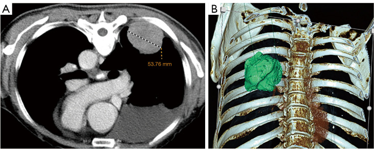
Figure 1.
First enhanced chest CT. (A) The cross-section of contrast-enhanced chest CT in the prone position showed a solitary mass near the chest wall in the right lower pulmonary lobe. The maximum diameter of the mass was 53.76 mm. The mass was slightly and uniformly enhanced after enhancement, and a medium amount of effusion was seen in the right thoracic cavity; (B) three-dimensional reconstruction intuitively showed the relative relationship between the tumor in the right lower pulmonary lobe, the ribs, and the spine. CT, computed tomography.

