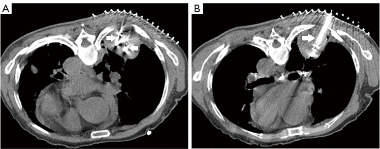
Figure 2.
Needle CT images of the tumor in the right lower pulmonary lobe during the first MWA. Double-point ablation was adopted in the area below the tumor (A) and single-point ablation was adopted in the area above the tumor (B). The white arrow points to MWA needle. CT, computed tomography; MWA, microwave ablation.

