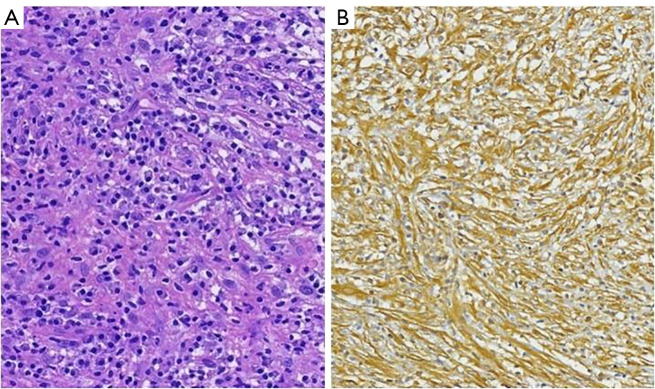
Figure 3.
Pathological examination reconfirming pulmonary IMT. (A) Histological examination showed a spindle cell tumor with infiltration of lymphatic and plasma cells. Hematoxylin and eosin staining, original magnification ×100. (B) Immunohistochemistry showed Vimentin positive, original magnification ×100. IMT, inflammatory myofibroblastic tumor.

