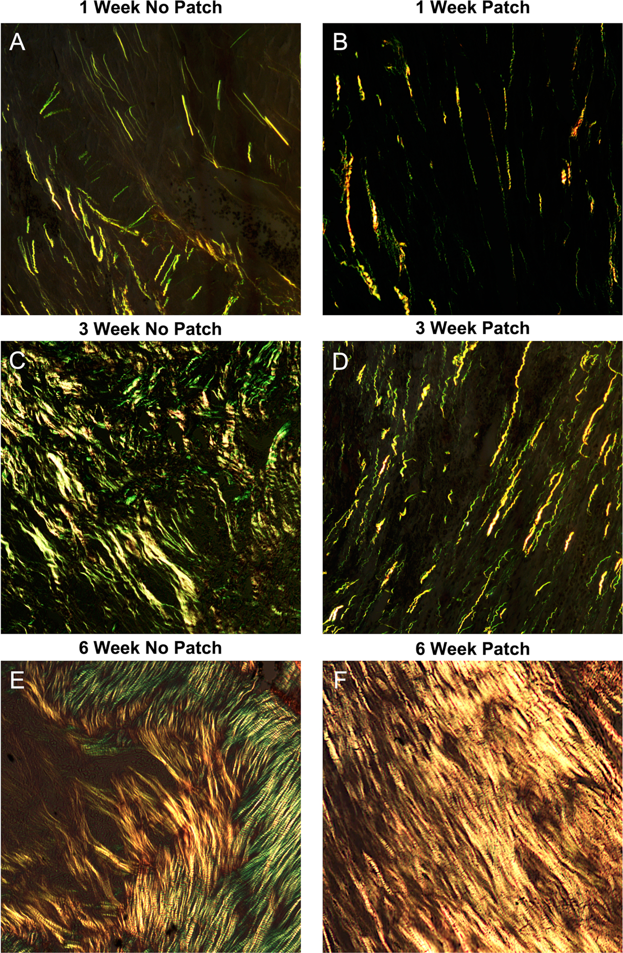
Fig. 1.

Representative images of picrosirius red-stained scar samples 1, 3, and 6 weeks after myocardial infarction. A,C,E: In the group with no patch, scars had dense, straight fibers with no preferred direction of collagen alignment. B,D,F: In the patch group, collagen had similar density at each time point but with a clear and strong alignment
