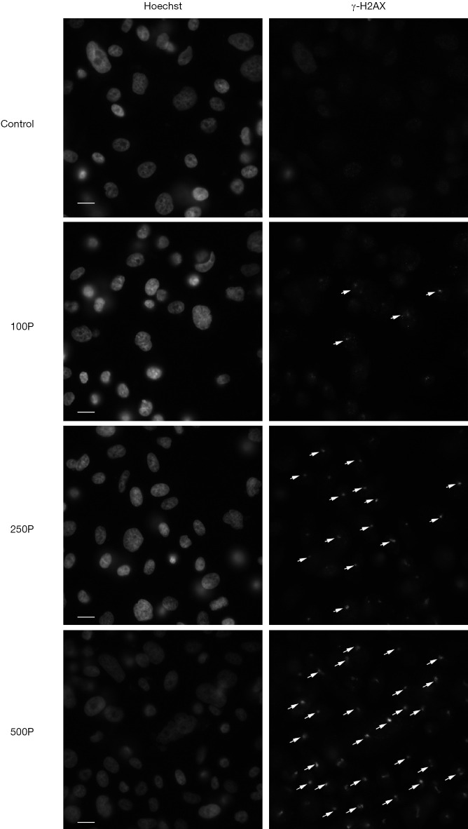
Figure 4.
Representative image of γ-H2AX expression level after irradiated by different number of protons. Cells were irradiated with 100, 250 and 500 protons. The bright-field illumination indicates DSB. The white arrow sign indicates γ-H2AX foci. The left photos were stained by Hoechst 33342, and the right-side photos were stained by γ-H2AX antibodies. Scale bar: 20 µm. DSB, DNA double-strand breaks.

