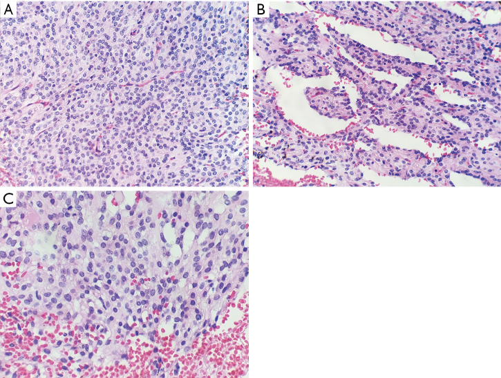
Figure 2.
Histopathological features (H&E). (A) Solid cellular nests and sheets of cells with an epithelioid appearance (400× magnification); (B) pseudopapillary structures (400× magnification); (C) the tumor cells containing round to oval nuclei with finely dispersed chromatin. Some tumor cells had vacuolated cytoplasm (400× magnification).

