Figure 3.
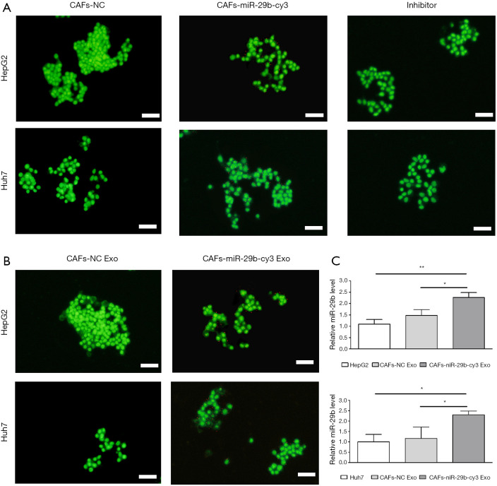
Transfer of miR-29b from CAFs to HepG2 and Huh7 cells via exosomes. (A) CAFs transfected with Cy3-labeled miR-29b (CAF-miR-29b-Cy3) or untransfected control cells (CAF-NC) were cocultured with GFP-labeled HepG2 and Huh7 cells for 24 h. The inhibitor group was exposed to 10 µM of GW4869. The red and green fluorescence signals were detected in HepG2 and Huh7 cells. Scale bar, 100 µm. (B) HepG2 and Huh7 cells were incubated with the exosomes (200 µg) extracted from CAF-conditioned media with Cy3-labeled miR-29b (CAF-miR-29b-Cy3 exo) or without (CAF-NC exo) transfection for 24 h. Scale bar, 100 µm. (C) qRT-PCR analysis of miR-29b expression in HepG2 and Huh7 cells exposed to CAF-derived exosomes (200 µg) transfected with pre-miR-29b-containing lentiviral plasmids (CAF-miR-29b exo) or NC group (CAF-NC exo). Representative images are shown. All data are presented as means ± standard deviations. *, P<0.05 and **, P<0.01 compared with each group. CAF, cancer-associated stromal fibroblast.

