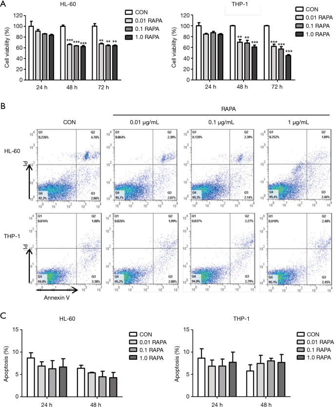
Figure 1.
Anti-leukemia effect of RAPA on AML cells. (A) Proliferation inhibition efficiency of various concentrations of RAPA (0, 0.01, 0.1, 1.0 µg/mL) on AML cells (HL-60 and THP-1) for 24, 48, and 72 h. AML cells were incubated in 96-well plates at a density of 1×10⁴ cells per well for 24, 48, and 72 h, and then CCK-8 solution was added for a further 4 h incubation. Results are reported as mean ± SD of the 3 independent experiments. Statistical significance was reported as **, P<0.01, ***, P<0.001; (B) HL-60 and THP-1 cells treated with different doses of RAPA (0, 0.01, 0.1, 1.0 µg/mL) were analyzed by FACS for the percentage of apoptotic cells for 48 h. Q1, dead cells; Q2, advanced apoptosis cells; Q3, living cells; Q4, early apoptosis cells; (C) cell apoptosis of AML cells (HL-60 and THP-1) following RAPA treatment. Each column and error bar represent the mean ± SD of apoptosis level (n≥2). RAPA, rapamycin; AML, acute myeloid leukemia; SD, standard deviation.

