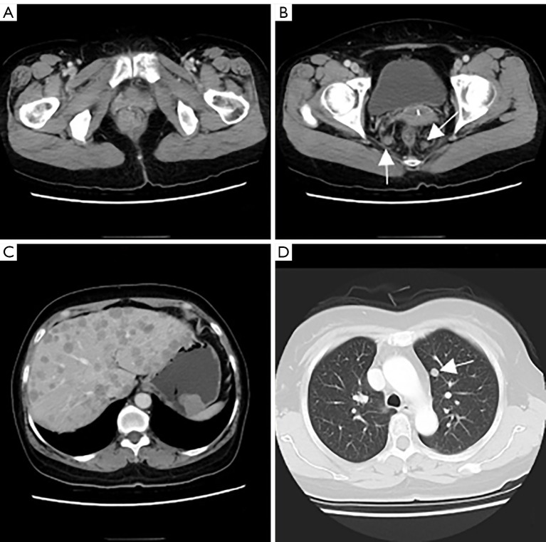
Figure 1.
CT of chest and abdomen. (A) Mass inside and outside of the lower rectum that showed obviously enhancement; (B) enlarged mesenteric lymph nodes were dispersed around (arrows); (C) protruded mass in the gastric fundus which also showed obviously enhancement, multiple metastatic lesions of liver; (D) small nodule in the left upper lung (arrow). CT, computed tomography.

