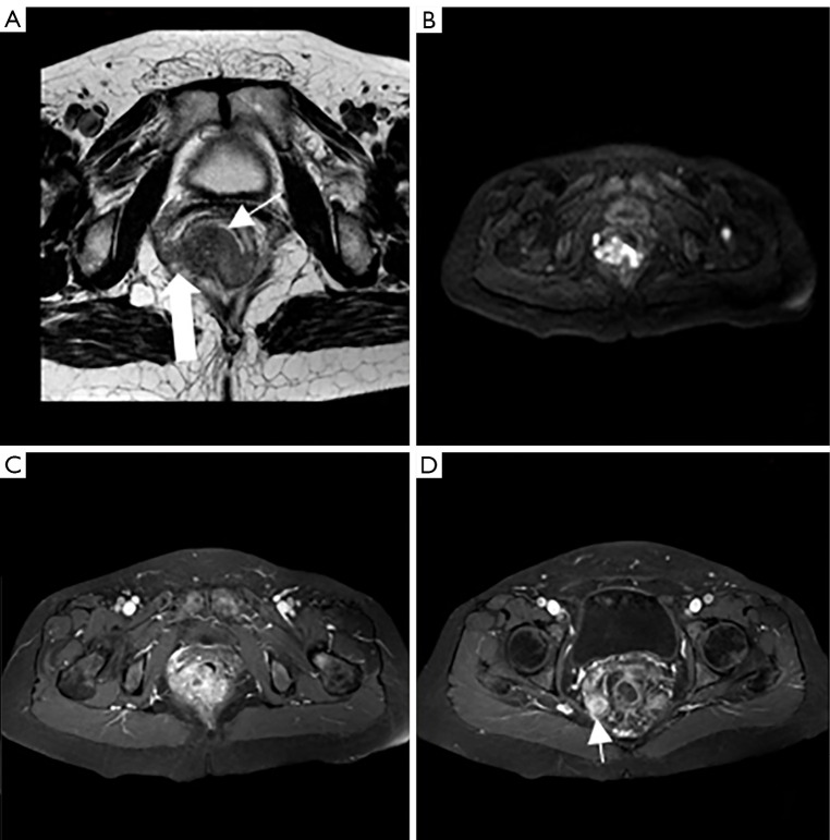
Figure 2.
Pelvic MRI. (A) Submucosal mass in the lower rectum, with continuous mucosal membrane (thin arrow) but involvement of the outer membrane and adjacent fat tissue (thick arrow); (B) diffusion-weighted-imaging presented high and uneven signal; (C) the mass was obviously uneven enhancement; (D) the mesenteric lymph node showed circular enhancement (arrow). MRI, magnetic resonance imaging.

