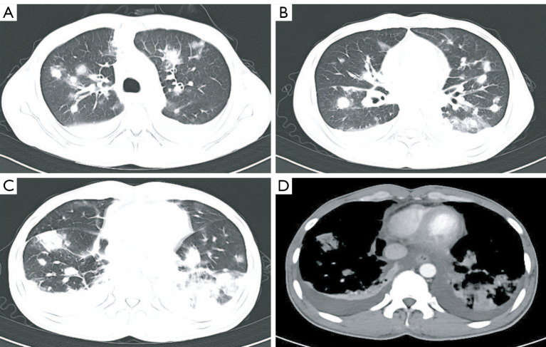
Figure 1.
Computed tomography images showing radiological changes. (A-C) The lung window photography of CT showed multiple nodules with ground-glass opacities and consolidation in bilateral pulmonary, thickening of bronchial walls, and interstitial pulmonary edema. (D) The mediastinal window photography of CT showed Mediastinal lymphadenopathy in the pretracheal retrocaval region and pleural effusion accompanied by inadequate expansion of both lower lungs.

