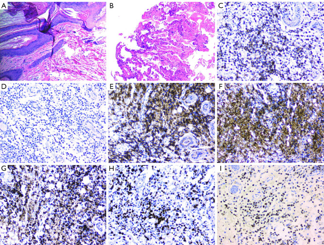
Figure 3.
Pathologic findings of Axillary mass and CT-guided transthoracic needle biopsy. (A) Tissue showed Malignant tumor with necrosis, tumor invasion of blood vessel wall and skin accessories, nerve involvement. (B) Percutaneous transthoracic needle biopsy specimen showed chronic inflammation, diffuse alveolar cavity visible large degenerative necrotic exudates and alveolar-epithelial atypical hyperplasia. (C) By immunohistochemistry, cells were positive for CD3. (D) Immunohistochemical staining was negative for CD20. (E) Immunohistochemical staining was positive for CD56. (F-G) Immunohistochemical staining was positive for granzyme B and TIA. (H) cells were positive for Ki-67. (I) In situ hybridization for Epstein-Barr virus-encoded RNA (EBER) showed positive reaction in tumor cells (magnification, ×100).

