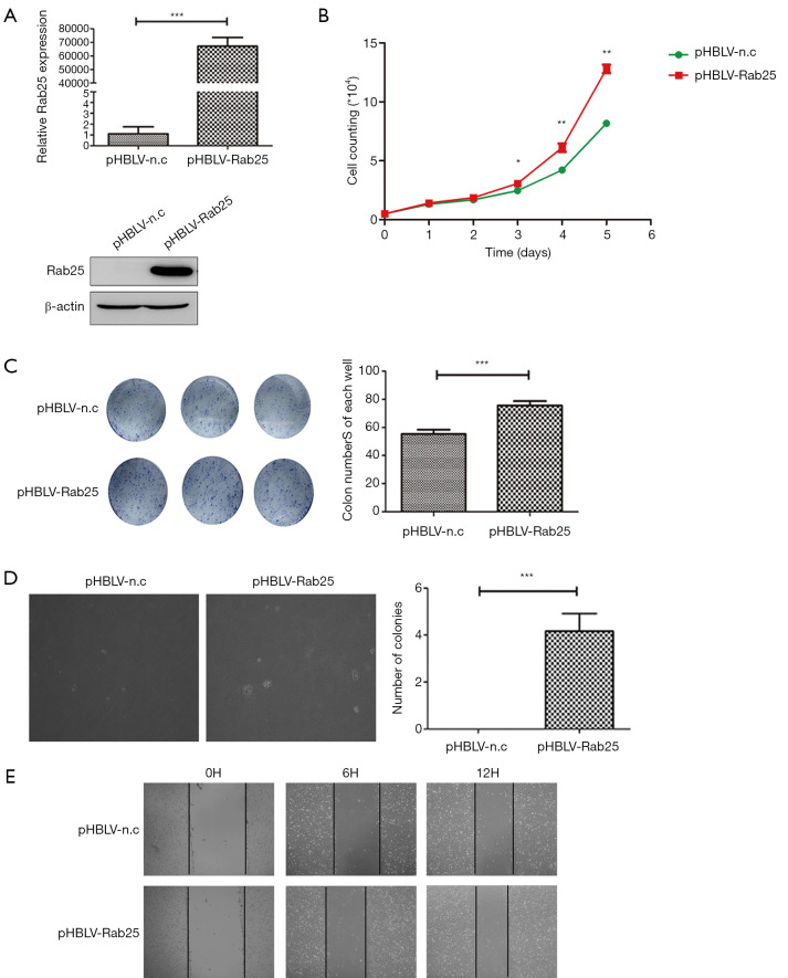
Figure 3.
Rab25 up-regulation in GES-1 cells promotes cell proliferation and migration. (A) qPCR analysis (normalized to β-actin) and western blot analysis (normalized to β-actin) of Rab25 expression in GES-1 cells transfected with pHBLV-n.c or pHBLV- Rab25 lentivirus. ***, P<0.001. (B) The growth curves of the pHBLV-n.c and pHBLV-Rab25 GES-1 cells by survival cell count assay in vitro. *, P<0.05; **, P<0.01. (C) Representative images (left) and quantification (right) of colony formation in six-well plates from the Rab25 overexpression GES-1 sublines. ***, P<0.001. Stained by coomassie brilliant blue dye and clones in 6-well plate without magnification. (D) Representative images (left) and quantification (right) of colony formation per 10-fold field of view in soft agar from the pHBLV-n.c and pHBLV-Rab25 GES-1 cells. ***, P<0.001. (E) Cell migration was detected using wound healing assay to investigate the effect of Rab25 in GES-1 cells. Photographs were taken at 0, 6, and 12 hours. Stained and magnified ten times.

