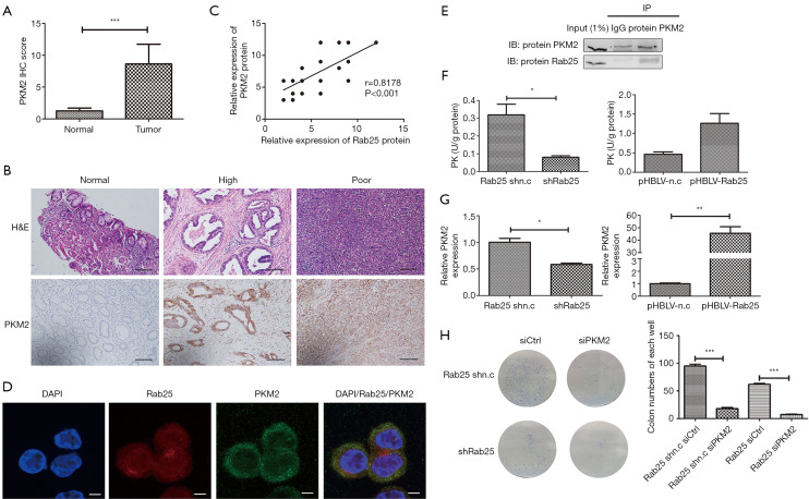
Figure 5.
Rab25 is a positive regulator of PKM2. (A) Comparison of IHC quantification of PKM2 expression from human gastric adenocarcinoma samples and matched normal gastric tissue samples (n=41). ***, P<0.001. (B) IHC staining of PKM2 proteins in human normal gastric tissues versus in human gastric adenocarcinoma samples. Stained by hematoxylin staining solution and scale bars were 200 µm. (C) Correlation analysis between PKM2 and Rab25 protein expression. n=41(Some points overlap), R=0.8178, P<0.001. (D) Immunofluorescence imaging of the expression and localization of intracellular Rab25 (red), PKM2 (green), and the nucleus (DAPI, blue) in the AGS cells. Stained by fluorescence and scale bars were 10 µm. (E) Rab25 interacts with PKM2 under physiological conditions in AGS cells. AGS cell lysates were immunoprecipitated with mouse monoclonal anti-PKM2 or mouse IgG. The immunoprecipitates were analyzed by western blots with anti-PKM2 (top) or anti-Rab25 (bottom) antibody. Input: positive control. (F) Pyruvate kinase (PK) activity measured in Rab25 knockdown AGS cells and Rab25 overexpression GES-1 cells. *, P<0.05. (G) The mRNA expression of PKM2 (normalized to β-actin) in Rab25 knockdown AGS cells and Rab25 overexpression GES-1 cells by qPCR analysis. *, P<0.05; **, P<0.01. (H) Representative images (left) and quantification (right) of colony formation in six-well plates from the siCtrl and siPKM2 AGS stably transfected cell lines. ***, P<0.001. Stained by coomassie brilliant blue dye and clones in 6-well plate without magnification.

