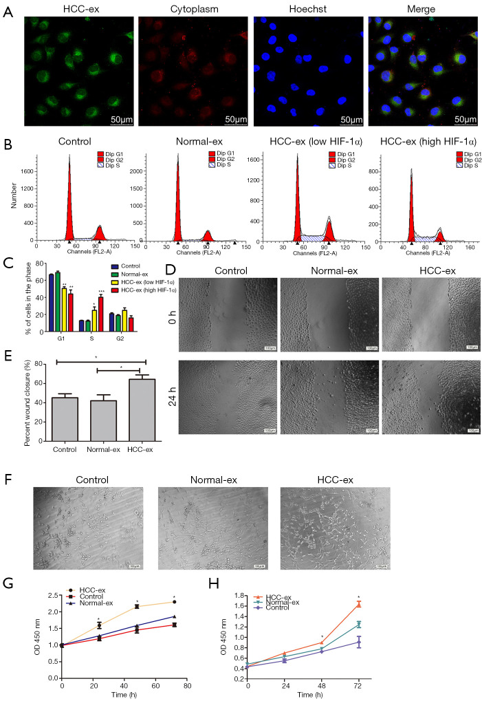
Figure 2.
HCC-exosomes promote tumor angiogenesis in vitro. (A) HUVECs were cultured with PKH67-labeled exosomes derived from HCC patients. The signals were detected in PKH67-labeled exosomes (green), the cytoplasm of HUVECs (red) and via nuclear counterstaining (blue). (B) Flow cytometry was conducted to (C) analyze the cell cycle in HUVEC cells. (D and E) Migration of HUVEC cells was detected by wound-healing assay following culture with exosomes or control. (F) HUVECs were cultured for 24 h in the absence (PBS) or presence of normal and HCC exosomes and then grown on Matrigel. Representative photomicrographs of tubes from the different treatment groups was presented. (G and H) Proliferation analysis was performed following exosomes being combined with (G) HUVECs or (H) Huh-7 cells. Data were expressed as the mean ± standard deviation. *P<0.05. HCC, hepatocellular carcinoma; HIF, hypoxia inducible factor; HCC-ex, exosomes derived from HCC patients; Normal-ex, exosomes derived from healthy individuals; HCC-ex (low HIF-1α), exosomes derived from HCC patients with a low level of HIF-1α protein; HCC-ex (high HIF-1α), exosomes derived from HCC patients with a high level of HIF-1α protein. HCC, hepatocellular carcinoma; HUVEC, human umbilical endothelial cell.

