Figure.

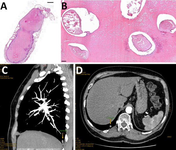
Histologic examination of resected tissue from a 66-year-old woman from southwestern Slovakia. A, B) Cross section showing Dirofilaria immitis nematodes embedded in necrotic material obtained from well-defined pulmonary nodule. Hematoxylin and eosin staining; original magnification ×20 for panel A, ×100 for panel. C, D) Chest computed tomography scan showing a subpleural focal lesion in the S10 segment of the right lung (arrows).
