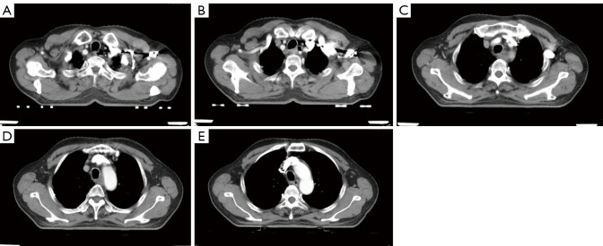
Figure 1.
A 68-year-old male presented with middle and upper esophageal cancer. The chest-enhanced computed tomography (CT) imaging showed thickening of the middle and upper esophageal wall, of approximately 1.7 cm, and stenosis of the lumen (A,B). After enhancement, there was mild to moderate inhomogeneous continuous enhancement (C-E).

