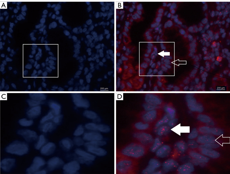
Figure 1.
Representative images of telomere fluorescence in situ hybridization in different tissue types. (C) and (D) were the partial enlarged drawing of (A) and (B), respectively. (A) and (C), nuclei were counterstained with DAPI (blue florescence). (B) and (D). Telomere length was reflected by the total red fluorescence intensity in nuclei. In CRC tissue, the CAF cells (filled arrows) show bigger, more numerous and intense red signal red signal than carcinoma cells (open arrows), reflecting longer telomere length in CAF. CAF, cancer-associated fibroblast cell.

