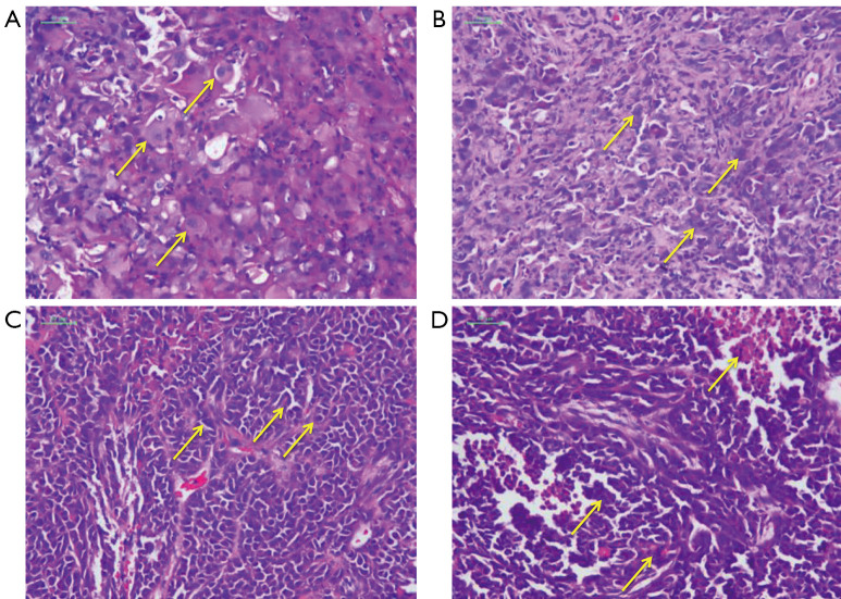
Figure 5.
HE staining of tumor tissue slices from mice (×200). (A) H226 control. Arrow indicating morphologically intact tumor cells with large cell volume, large and distinct nucleoli; (B) H226 Maytenus compound. Arrow indicating degenerative changes tumor cells with small cell volume, concentrated nucleus and plasma; (C) HeLa control. Arrow indicating morphologically intact tumor cells with large cell volume, large and distinct nucleoli; (D) HeLa Maytenus compound. Arrow indicating degenerative changes and necrotic tumor cells with small cell volume, concentrated nucleus and plasma.

